
A Transabdominal Sonography (TAS) ultrasound uterus exam is performed using a transabdominal 3.5 MHz transducer. Initially, a standard gray scale, three-dimensional, and then the Doppler ultrasonography Transabdominal ultrasonography is going to be performed with the patient in dorsal decubitus and with a full urinary bladder. On the other hand, the Trans-vaginal sonography TVS is going to be performed using a - MHz transducer. The probe is cleaned with spirit using cotton first before it is inserted after which it is covered with ultrasound gel and condom. Once the scan is completed, the probe is again thoroughly cleaned and wiped with spirit.
The most significant landmark of pelvic anatomy is the uterus. It is found in mid-centre of pelvis. It is very rare to find a laterally positioned uterus, but it is found around the axis of the uterine isthmus. During the measurement of the size of the uterus, there are several measurements that are required for the ultrasound. One of these is length, which refers to the distance from the fundus to the internal orifice of the uterus on a sagittal view, the width, which is largest anterior-posterior distance measured in the mid portion of the uterine body also on a sagittal view, the depth which refers to the maximum distance measured at the level of the uterine fundus on a transverse view. Uterine isthmus is identified where the uterine body and cervix meet.
Additionally, the endometrial thickness is measured using the sagittal view. When the cavity line is present the endometrial thickness is measured from the base of its anterior layer to the base of its posterior layer double layer at proximal, middle and distal uterus in the sagittal plane. The thickness and the echogenicity of the endometrium vary with the different stages of the menstrual cycle. The myometrium, in this case, is defined as a layer of homogeneous echogenicity from the serosal surface to the decidual. It has a structure, with fine, parallel, linear echoes and an echogenicity similar to a muscle
When it comes to the Focal lesions of myometrium and cervix there are several etiologies that are mostly reported for abnormal uterine bleeding diagnosed on imaging. They include leiomyomas, adenomyosis, endometrial hyperplasia, and endometrial polyps.
Leiomyomas: They are the most common pelvic tumors, occurring in up to 25% of women over the age of 35.Leiomyomas are composed of smooth muscle cells arranged in a whorl-like pattern with variable amounts of intervening collagen, extracellular matrix, and fibrous tissue. The initial imaging study
Adenomyosis; Adenomyosis is a condition in which ectopic endometrial glands and stroma are embedded in the myometrium. Adenomyosis is seen in 19% to 62% of hysterectomy specimens, often presenting symptomatically in the reproductive years and within the perimenopausal years.
Endometrial hyperplasia: The excessive proliferation of the endometrial glands is termed endometrial hyperplasia. The endometrial gland-to-stroma ratio is increased and the glands can be cystically dilated.
Endometrial polyps: Endometrial polyps are focal excrescences of endometrial hyperplasia covered by endometrium. Three types of endometrial polyps exist: hyperplastic, atrophic, and functional.
Research Method
The research method used in this case was quantitative research method, with the results being determined through the use of the measurement devices mentioned in the paper. A cross-sectional study was conducted in this case to determine the most accurate method between the transabdominal sonography TAS and transvaginal sonography TVS in perimenopausal abnormal uterine bleeding patients. This comparative study was carried out in a hospital in the Ultrasound Section, Department of Radiodiagnosis and Imaging. Additionally, an independent observer was used in the comparison of the results.
Results
The average results for the endometrial thickness by TVS and TAS are shown in the table below.
Endometrial thickness TAS (Average) Endometrial thickness TVS (Average)
Proximal Middle Lower proximal Middle Lower
0.74 0.78 0.62 0.77 0.90 0.80
Myometrial thickness TAS (Average) Myometrial thickness TVS (Average)
Anterior Posterior Anterior Posterior
1.23 1.28 1.65 1.55
The results for the patients with adenomyosis (Menorrhagia) were compared for the endometrial thickness. The endometrial thickness for the patients with adenomyosis is shown below. The age of the patien,t in this case, was 56 years.
Endometrial thickness TAS Endometrial thickness TVS
Proximal Middle Lower Proximal Middle Lower
5.5 3.8 3.9 5 4 3.9
When it came to the adenomyosis for Oligomenorrhea, the patient was 45 years old. The endometrial thickness comparison is shown in the table below.
Endometrial thickness TAS Endometrial thickness TVS
Proximal Middle Lower Proximal Middle Lower
0.3 0.2 0.1 0.3 0.3 0.3
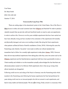
The results obtained above are also shown in the chart on the next page.
Figure SEQ Figure \* ARABIC 1Endometrial thickness comparison for the 45 year old patient
TVS is represented by the green color
TAS Is represented by the Maroon color
For the patients with the endometrial polyp, the endometrial thickness for Menorrhea patients was not carried out for the patients who were 48years old. For the patients who were who were 43 years old, a comparison of the endometrial thickness is shown below.
Endometrial thickness TAS Endometrial thickness TVS
Proximal Middle Lower Proximal Middle ;Lower
0.23 0.34 0.34 0.3 0.34 0.34
For the menorrhea case, the patient was 52 years old. The endometrial thickness measurement is shown below for the patients with the endometric polyp.
The average measurement for the Oligomenorrhea, for the patients with endometrial polyp, is shown below. The age of the patient was 52 years old.
Endometrial thickness TAS Endometrial thickness TVS
Proximal Middle Lower Proximal Middle Lower
0.21 0.06 0.3 0.14 1 0.36
Additionally, the average endometrial thickness measurement classified in age groups from 40- 45, 46-50 and 51-55 years is shown in the table below.
Age group Endometrial thickness TAS Endometrial thickness TVS
Proximal Middle Lower Proximal Middle Lower
40-45 0.64 0.67 0.59 0.62 0.82 0.85
46-50 0.82 0.98 0.57 1.13 1.13 0.71
51-55 0.87 0.81 0.78 0.72 0.83 0.80
Discussion
Looking at the age of all the participants of the study, it is seen that their age ranges from 41 to 66 years. Their mean age is 47 years. When the uterine length is compared between the TAS and TVS, it is seen that the TAS uterine length is considerably longer than the length by TVS. The average uterine length by TAS is found to be 8.67 while that of TVS is 7.5 mm. When it comes to the uterine width, it is the complete opposite as the uterine width by TAS is less than that by TVS. The average width by TAS is 4.4 while that by TVS is 4.6. For the height, they are almost equal to each other as the average height by TAS is 4.64and the average height by TVS is 4.66. It can, therefore, be concluded that the major measurement that showed a great and significant variation was the length of the uterine length. As seen from the resulted for both TAS and TVS, it is clear that TAS showed the criteria for a bulky uterus. However, TVS length never reached the criteria for a bulky uterus. Therefore, it can be concluded that TAS is superior over TVS to determine bulky uterus.
The proximal endometrial thickness by TAS was compared with that for TVS. The average value for the TAS measurement was 0.74 while that by TVS was 0.77. When it came to the middle endometrial thickness by TAS, the average was 0.78 while that by TVS was 0.9. Hence, the middle measurement by TVS was also higher than that by TAS. In the lower endometrial thickness measurement by TVS still had a larger measurement. This is because, on calculation of the average measurement by TVS, the value was 0.804 while that by TAS was 0.62.
The myometrial thickness measurement by TAS was found to be less than that by TVS. This refers to the anterior myometrial thickness. The average number of the anterior myometrial thickness by TAS was approximately 1.28 while the average by TVS was approximately 1.65. When it came to the posterior myometrial thickness, the value by the TAS was also less than that of the value by TVS. The average by TAS was 1.28 with the average by TVS standing at 1.55. In all the cases mentioned above, the average number of participants in all the scans was 59 patients. All of the subjects participated in the scans for every condition. When using TAS, some of the results obtained for some patients indicated that there was the presence of polyps. However, it was concluded that to be submucosal fibroid from the TVS result. This showed that submucosal fibroid was visible when using TVS. However, there was no difference between TVS and TAS when it came to the intramural fibroid. In some cases, abnormal uterine bleeding patients had an unexpected finding of an intrauterine collection which is evident in the 46-50 age groups mostly. The most probable cause of this observation is due to stasis of blood clots for a long duration of time or proximal to the endometrial polyp especially in patients with prolapsed polyps or submucosal fibroids.
In some patients, there was the detection of adenomyosis when TVS was used. This was not exhibited in the TAS detection. There is a possibility that adenomyosis and endometrial cysts are related because, in the test, the two conditions were detected hand in hand. A patient with endometrial cysts also had adenomyosis. Hence, it was concluded that both of the two were related and a patient could not have one without the other.
In a gynecological evaluation, it is required to have a meticulous uterine cavity evaluation. This is required for all the situations that can arise such as non-bleeding symptoms of the condition of the uterine, abnormal uterine bleeding, and all the other cases that may arise. One of the biggest gynecological issues that face women is the Abnormal uterine bleeding. Hence, there are several methods that are available for conducting the investigations, among them TAS and TVS. It is therefore important to have an idea on which is the best method for conducting the evaluation due to the availability of several options. From the analysis of the results obtained from this study, it can be concluded that Transabdominal sonography is not as effective as transvaginal sonography when it comes to large pelvic masses. However, TAS is superior to TVS in other cases that range from the endometrial carcinoma, polycystic ovaries, ectopic pregnancy, and many other cases. For the cases that involve patients with inflammatory disease of the pelvis that is advanced, there is no visible difference in their accuracy. Hence, they can be both said to be suitable for such cases.
When it comes to the pelvic pathology, TVS is better and more accurate than TAS. Despite this, the Transabdominal sonography should be the one that is used as the initial monitoring method when investigating the condition of the female pelvis.
Request Removal
If you are the original author of this essay and no longer wish to have it published on the customtermpaperwriting.org website, please click below to request its removal:

